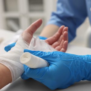
Air embolisms are caused when gas bubbles enter blood vessels. This results in poor blood flow and decreased oxygen delivery to the areas where the blood vessels have been affected. Air embolisms can be fatal or result in serious disabilities. Following an air embolism a person might experience weakness or paralysis in the limbs, loss of vision, heart, lung or brain damage and many other permanent health conditions. Aggressive treatment of air embolism is needed to ensure the best chance of recovery from this disease.
Hyperbaric oxygen therapy has been used to reduce the size of bubbles circulating in blood vessels.
The increased pressure in the hyperbaric chamber makes the bubbles smaller and helps push them back into physical solution, while the high oxygen pressure washes out the gas from the bubble. Once the bubbles are smaller or gone, blood flow resumes.
This allows poorly oxygenated tissues to receive high levels of oxygen. Also, when vessels are obstructed by gas bubbles they leak fluid causing swelling in the surrounding tissues. When blood flow is restored, the swelling subsides, improving blood supply and oxygen delivery even more. Lastly, high amounts of oxygen provided in the hyperbaric chamber support the areas injured by air embolisms while blood flow reduction and tissue swelling are being repaired. Air embolisms are medical emergencies and are treated within a hospital setting.
HBOT can help with the effects of spider bites.
This type of injury has been shown to respond to hyperbaric oxygenation treatments. Necrotizing (flesh eating) effects are a complication of the brown recluse spider and other spider bites which are characterized by progressive necrosis and inflammation of the tissue at the site of the spider bite. Doctors sometimes diagnose this by the signs and symptoms that later develop. How much tissue damage you get is relative to the amount of venom injected, location of the bite (with high fat areas being more severely affected) and the immune status of the patient.
I remember a patient we had several years ago. They ended up in the emergency room with 6 bites on the their lower left leg. Each site had a width of 3 inches of tissue destruction. The plastic surgeon had told the patient that they may have to amputate the leg. Fortunately, the doctor referred the patient to Northern Nevada Hyperbarics and after two weeks of treatment (10 days) the patient was on the road to recovery and the wounds healed nicely.
If you or a loved one ever get into a situation where you get a spider bit and it is not healing get to a doctor immediately. It is a very serious situation. Contact us at (775) 826-2084 if you have any questions. We are here to help you.
As we approach the winter time in the Truckee Meadows, please consider how to protect you and your family from Carbon Monoxide Poisoning. Carbon Monoxide Poisoning is a leading cause of death by poisoning in the United States. Among causes of Carbon Monoxide Poisoning are automobile exhaust (accidental or purposeful), faulty heaters, and building fires. Hyperbaric Oxygen Therapy is an approved treatment for this serious condition.
Many factors affect the carbon monoxide poisoning symptoms and the outcome of treatment.
Most significant factors are: the inhaled CO concentration, duration of exposure, rate and depth of breathing, heart rate, co-morbid illnesses and most importantly, the time between discovery of the patient after exposure and arrival at a Hyperbaric Chamber.
Carbon Monoxide Poisoning
Stephen R. Thom, M.D., Ph.D., FACEP and Lindell K. Weaver, M.D., FACP, FCCP, FCCM
The injuries caused by carbon monoxide (CO) traditionally have been viewed as due to a hypoxic stress mediated by an elevated carboxyhemoglobin (COHb) level. The two organ systems most susceptible to injury from CO are the cardiovascular and central nervous systems. Human and animal data indicate that major cardiac injury is due primarily to CO-induced hypoxic stress.
However, the COHb level does not correlate well with the development of neurological injuries. Recent investigations have established that systemic oxidative stress can arise from exposure to CO and that perivascular and neuronal injuries arise by mechanisms other than hypoxia. Neuropathology seems to be due to a complex cascade of biochemical events involving several pathophysiologic processes.
Administration of supplemental oxygen is the cornerstone of treatment of CO poisoning. Oxygen inhalation will hasten disassociation of CO from hemoglobin dissociation to occur at a rate greater than that achievable by breathing pure oxygen at sea-level pressure.
Additionally, HBO2, but not ambient pressure oxygen treatment, has several actions which have been demonstrated in animal models to be beneficial in ameliorating pathophysiologic events associated with central nervous system (CNS) injuries mediated by CO.
These include an improvement in mitochondrial oxidative processes, inhibition of lipid peroxidation, and impairment of leukocyte adhesion to injured microvasculature. Animals poisoned with CO and treated with HBO2 have been found to have more rapid improvement in cardiovascular status, lower mortality, and lower incidence of neurological sequelae.
Since 1960, the clinical use of HBO2 for CO poisoning has occurred with increasing frequency. Over 2,500 CO-intoxicated patients were treated in North American hyperbaric chambers in 1992. However, this is only a small fraction of those poisoned with CO. Extrapolation of data from a 1994 survey across three western states projected that over 4,000 CO-poisoned patients are evaluated in emergency departments annually in the United States.
In reported series, clinical recovery among patients treated with HBO2 appears to be improved beyond that expected with ambient pressure supplemental oxygen therapy. This has been observed both in terms of mortality and neurologic morbidity.
This research found that the optimal benefit from HBO2 occurs in those treated with the delay after exposure and that repeat treatments may yield a better outcome than just a single treatment in selected patients.121
I have personally treated numerous patients over the last 11 years with non healing wounds. (Check out the graphic images below) Sometimes, people were one step away from amputation. It is so rewarding to save toes, feet, and whole legs (and other body parts) and to see how we can give back a quality of life that our patients never thought possible.
Do you or someone you know have a non healing wound that stems from vascular insufficiency, a complication after radiation therapy, diabetes, or a problem amputation site? How about a wound that comes from a traumatic injury or one that just won’t heal after surgery?
These wounds could all share the common underlying problem of having a low oxygen level, what doctors call tissue hypoxia usually related to poor circulation. Add to the common problem of low oxygen, tissue inflammation and/or infection and you have the ingredients for what could become a very serious limb or life threatening situation.
Sometimes wounds fail to respond to wound care alone. This happens especially when a person has multiple complicating factors like diabetes or a history of radiation therapy.
Fortunately, when hyperbaric oxygen treatment is used in conjunction with standard wound care, patient outcomes improve substantially. Give us a call and/or tell your doctor that you would like to try hyperbaric oxygen therapy. Contact Richard Flyer, Clinical Director at (775) 826-2084. We offer a free consultation.
Key to Images:
First Row—-Diabetic wound with a bone infection. Healed after 35 treatments (7 weeks)
Second Row—Crush injury to hand. 3 treatments to start for a total of 20 total (4 weeks)
Third Row—-Wound from radiation to the neck. 35 treatments (7 weeks)
Fourth Row—Open wound on knee from radiation. Needed 20 treatments (4 weeks)-graft applied after.
A skin graft is a transplanted tissue without its blood supply which is transferred during skin grafting surgery, a type of surgical grafting where transplantation of skin is performed. A graft consists of a section of skin including epidermis and dermis of variable thickness that has been completely separated from its blood supply and transplanted to a recipient bed.
Reconstructing complex wounds is accomplished by shifting or transferring tissues to the wound from a different part of the body. The area from where the skin is borrowed is called the donor site.
Skin Grafts Categories
There are two general categories of grafts in regards to a thickness: full thickness and partial thickness grafts. A full thickness grafts contains the dermis and the epidermis. A partial thickness skin graft includes the epidermis and only a small portion of the dermis. The split thickness skin grafts range from 1/1,200 to 1/2,000 of an inch in depth. Thicker grafts are less prone to wound contracture. In general, full thickness grafts are used in places where less wound contraction is desired, such as the face.
Skin grafts survive as oxygen and nutrients diffuse into them from the underlying wound bed. Long-term survival depends on a new blood supply forming from the wound to the graft. When the wound bed does not have enough oxygen supplied to it, the skin graft will at least partially fail.
Common causes for this are previous radiation to the wound area, diabetes mellitus, and certain infections. In these situations, the availability of oxygen in the wound bed can be increased with hyperbaric oxygen therapy (HBO2) in preparation for skin grafting. Additionally, HBO2 can be used after skin grafting to increase the amount of the graft that will survive in these compromised settings.
Skin Grafts – Hyperbaric Oxygen Therapy, Skin BurnsOnce the graft has been harvested, it becomes separated from its blood supply and must revascularize to survive. The vascular bed is recreated in a three-step process: imbibition, inosculation, and revascularization. Imbibition occurs in the first 24 to 48 hours. During this phase, the nutrients diffuse from the underlying wound bed to extracellular fluid and into the capillaries of the graft.
The graft begins to fix to the wound bed via fibrin bonds. Inosculation involves the alignment of the donor vascular buds with graft capillaries and establishment of circulation. Revascularization occurs when connecting vessels differentiate into arterioles and venules. Thicker grafts demand more blood supply and require a well vascularized recipient bed to survive. Wounds with exposed bone, tendon, or cartilage or with radiation damaged tissue offer a poor vascular bed.
There are several reasons for graft failure. The most common cause is loss of contact with the recipient bed because of hematoma, seroma, purulent material, and shearing. Meshing the graft helps to overcome the problem of fluid buildup underneath it. Shearing is caused by movement of the graft against the wound bed. This can be prevented by tie-down compression dressings created by interrupted sutures along the edge of the wound. The ends of the suture are left long in order to tie down a nonadherent dressing on top of the graft. The second most common cause of graft failure is infection. Wounds colonized with more than 1 ª 105 bacteria per gram of tissue will be less likely to support a graft.
Partial thickness skin grafts are most commonly harvested with the use of a dermatome, which is air driven or electric. Dermatomes can be adjusted for depth of cut as well as width of the skin graft desired. Full thickness grafts are commonly acquired by hand with a scalpel.The incision is made in an elliptical fashion to facilitate wound closure. Securing the skin graft to the recipient bed can be accomplished using staples placed along the circumference of the graft. In addition, interrupted chromic sutures can be placed within the graft to anchor it to the wound bed.
Non-adherent dressing as well as sterile absorbent gauze are then placed, and the dressing is left on for 5 days for split thickness grafts and 7 to 10 days for a full thickness grafts. Healing of a split thickness donor site results from epithelialization from epidermal appendages, such as hair follicles, sweat glands, and sebaceous glands that are left behind after the graft is taken. Healing occurs best in an environment that is moist and free from injury and contamination. The process may take up to 21 days. Full thickness donor sites leave no dermis or epidermal appendages behind and, therefore, must be closed primarily or left open to granulate and contract.
Skin Flap
A skin flap differs from graft in that it is a section of tissue that comes with its original blood supply. The blood supply of the flap is referred to as the pedicle. There are several Types of skin flaps:
* Local flap. When the skin flap is from an area close to the wound, for example, a wound on the lip may be repaired by a flap from the adjacent cheek.
* Regional flap. When the skin flap is not from the adjacent area, but is from the same region of the body, for example, a wound on the tip of nose might be repaired with a flap from the forehead.
* Distant flap. When a flap is from a different part of the body, for example, a wound on the hand might be repaired with a flap raised in the groin. A local flap repair is usually done in one operation, whereas regional and distant flaps need two or more operations. The second operation is needed to detach one end of the flap at the donor site, when the blood vessels have developed at the other end.
* Free flap. This is a distant flap, but the whole procedure is done in one stage by repairing the donor and blood vessels by microsurgery.
Skin Flaps also require oxygen and nutrients to survive. The outer, visible portion (usually skin) is furthest from the source of blood supply for the flap. This is the area most likely to be compromised by inadequate oxygen.
Factors such as age, nutritional status, smoking, and previous radiation result in an unpredictable pattern of blood flow to the skin. If a flap is found to have less than adequate oxygen after it has been transferred, HBO2 can help to minimize the amount of tissue which does not survive and also reduce the need for repeat flap procedures.
Hyperbaric Oxygen Treatment is neither necessary nor recommended for the support of normal, uncompromised skin flaps and grafts. However, in tissue compromised by irradiation or other cases where there is decreased perfusion or hypoxia, HBO2 has been shown to be extremely useful in flap salvage. Hyperbaric oxygen can help maximize the viability of the compromised tissue thereby reducing the need for regrafting or repeat flap procedures. A number of studies have shown the efficacy of HBO2 on enhancement of flap and graft survival in a variety of experimental and clinical situations.
People in the old days just didn’t know the consequences of smoking.—
I thought I would share this story about a doctor who learned his mother was being treated with HBOT for radiation to her jaw. “I live a long ways from the rest of the family. I grew up….MORE
Modern cancer treatments utilize radiation therapy. Doses used to kill cancer growth can sometimes damage (5% of the time) the surrounding tissue. How radiation effects us is really an individual matter. These effects might be felt 6 months to years after the exposure.
Radiation can damage some of the blood vessels in the area where the cancer was. Having less blood supply and oxygen to those areas can lead to non-healing tissues.
Take cancer of the head and neck for example. The jawbone sometimes gets in the way of the path of radiation. The jawbone is very thick and hard and starts out with a smaller number of blood vessels compared to other tissues of the body. After radiation to the head and neck some small vessels feeding the jaw bone can be damaged.
Oral surgeons are concerned about what they call radiation carries or decay of teeth from radiation. In a jaw that has been exposed, the surgeon is concerned that removing damaged teeth could lead to complications such as infection or lead further still to actual death of part of the jawbone. So, HBOT is used prior to teeth extraction to help re-generate new blood vessels and following surgery to remove the affected teeth to help speed post surgical healing.
HBOT works on many other parts of the body–breast, prostate, bladder, gynocological area, GI, and abdomen.
Northern Nevada Hyperbarics has almost 11 years of treatment experience. We have the most experience with treating the effects of radiation therapy and other conditions such as non healing wounds. And, through our year old partnership with Renown Health, we have created the most modern facility where the quality of patient care is number one.
It’s that time of year, summer, for those of you who want to scuba dive at Lake Tahoe. It is a clear and pristine location to make that easy entry dive. Please remember that the lake is at 6400 ft. elevation and at present there is a lot of cold snow melt.
Keeping this in mind, you’ll need to prepare with high altitude dive tables and use a thick wet or dry suit. Also, think of how you get to the lake. Do you drive or fly there? Did you give yourself time to de-gas from sea level? Are you going to have to go up over a pass to get back to where you are staying? Do you know the symptoms of simple or severe decompression sickness? Include Northern Nevada Hyperbarics, the only facility treating decompression sickness, in your dive planning.
Northern Nevada Hyperbarics is at Renown Medical Center in Reno, NV. We are capable of treating all forms of decompression sickness. We have Sechrist mono place chambers with air-brake capabilities. Our staff has a combined 50+ years of experience.
Contact Frank Irwin (the smiling one in the photo above), safety and dive officer at (775) 826-2084
Copyright © 2012 Northern Nevada Hyperbarics Inc. · All rights reserved
Website Development and Design by Dan Marston & Integris Marketing
Powered by WordPress